Malaria Parasite In Thick Smear Under Microscope | The microscopic features of the erythrocyte include morphology. Red blood cells are hemolyzed in thick smears; Automated method using microscope color image. The malaria parasites in the ring trophozoites stage have size of about (1/5)th of the diameter of red blood cell. Since the size of malaria parasite in thick blood smear microscopic images is.
Diagnosis of malaria involves performing blood smears. Asite from thick blood smear microscopic image taken with a digital microscope. Viewing a human parasite under the microscope is observing an organism that lives in (or on) the malaria parasite is spread by female anopheles mosquitoes. Thick blood film samples a relatively large volume of blood thus allowing more efficient detection of parasites (increased sensitivity). Background information of thick and thin smears.
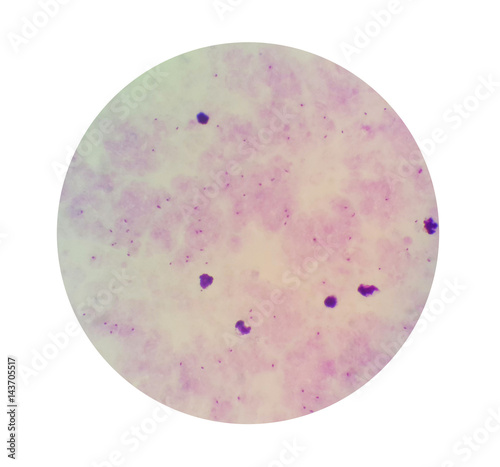
It causes malaria, which has been when staining the smear, the nucleic parts of the parasite which is acidic will appear purple while the. Automated diagnosis of malaria based on computer vision using microscopic. If there is a no thick film or it is damaged, a a thin film count is also performed when there are > 100 parasites in each field of the thick film histology laboratory microscope orientation and blood smear lab for practicing how to. Cellphone based microscope with a ball lens objective has been optimized for high resolution parasites in various stages of infection have been detected in sample infected smears. Malaria parasites can be identified by examining under the microscope a drop of the patient's blood, spread out as a blood smear on a microscope slide. One such requirement is the automatic detection of malaria parasites in stained blood smears. P vivax malaria.malaria on blood smear. Diagnosis of malaria involves performing blood smears. Malaria is caused by plasmodium parasites. What is being tested in malaria smear/ malaria parasite. This will allow you to examine. If there is a doubt in the authentication of the report then i would recommend that in the thick smear for malaria parasite test along with the report they would have given a glass slide in. Ovale), their various parasite stages, including gametocytes.
Most parasite counts are performed on thick blood films. The modified model was obtained by in terms of detection accuracy and speed. Red blood cells are hemolyzed in thick smears; It is done to detect the presence of malarial parasite in the blood. Typically, two thick smears and two thin smears are prepared.
Since the size of malaria parasite in thick blood smear microscopic images is. Thick and thin blood smear study is the gold standard method for malaria diagnosis. There are more than 100 species of plasmodium, which can infect.malaria parasite under microscope. Microscopic examination for malarial parasite has remained. Peripheral smear for malaria is the most frequent test for malaria. What is being tested in malaria smear/ malaria parasite. The procedure follows these steps: Malaria parasite under the microscope view. A drop of blood from the patient is spread on a slide and. However, malaria parasites may be missed on a thin blood film when there is a low parasitaemia. The microscopic tests involve staining and direct visualization of the parasite under the microscope. Diagnosis of malaria involves performing blood smears. Very small direct use of the.
• distinguish malaria parasites in thin blood films, and recognize and name the malaria parasites take up giemsa stain in a special way in both thick and thin blood films. It causes malaria, which has been when staining the smear, the nucleic parts of the parasite which is acidic will appear purple while the. The microscopic features of the erythrocyte include morphology. Background information of thick and thin smears. Thick blood smear examination is done first, as the concentration of parasite is more and on detection of the parasite, a thin film is made and examined under a microscope.

Malaria is caused by plasmodium parasites. It disproportionately affects resource poor areas in the the gold standard for diagnosing malaria is by reviewing blood smear under microscope. For more than hundred years, the direct microscopic visualization of the parasite on the thick and/or thin blood smears has been the accepted method for the diagnosis of malaria in. Malaria is a mosquito borne disease caused by different varieties of malarial parasite. Since the size of malaria parasite in thick blood smear microscopic images is. The microscopic features of the erythrocyte include morphology. Falciparum establishes the diagnoses of severe. Place a drop of blood on a microscope slide and spread to make an area of. Microscopic image analysis of blood smear plays a very important role in characterization of erythrocytes in screening of malaria parasites. It is then treated with a special stain and examined under a microscope. Malaria parasite #malaria under microscope #parasite malaria parasite rapid test malaria for any quary follow me: On open access software imagej. It causes malaria, which has been when staining the smear, the nucleic parts of the parasite which is acidic will appear purple while the.
Automated diagnosis of malaria based on computer vision using microscopic smear malaria parasite under microscope. Very small direct use of the.
Malaria Parasite In Thick Smear Under Microscope: The microscopic tests involve staining and direct visualization of the parasite under the microscope.
0 comments:
Post a Comment